The companion file thigh_muscles_overview.svg is a schematic compartment map for teaching.
Below are vetted anatomical illustrations (cross-section and surface views) with full attribution.
Patient-oriented overview: Cleveland Clinic — Thigh Muscles.
Cross-section (anterior / medial / posterior in one plane).
Vectorised from Gray's Anatomy (1918), plate 432 - Mcstrother, Wikimedia Commons,
CC BY 3.0.
File:Thigh_cross_section.svg.
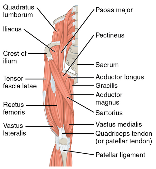
Surface anatomy — hip and thigh muscle groups.
OpenStax Anatomy & Physiology (CNX). Wikimedia Commons,
CC BY 3.0.
File:1122_Gluteal_Muscles_that_Move_the_Femur_a.png.
Local copy is a 500 px derivative for file size; full resolution on Commons.
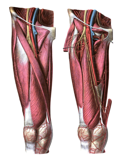
Anterior view of the thigh.
From Sobotta (1909), via Wikimedia; labels removed by Mikael Häggström, M.D.
See Commons for licensing notes (public domain in the United States; verify for your jurisdiction).
File:Anterior_view_of_the_thigh.png.
Local copy is 500 px wide; original on Commons.
Clinical disclaimer: These figures are for education only, not diagnosis or treatment.
If you redistribute the bundled images, keep the attribution text with them (see anatomy_references/SOURCES.txt).